
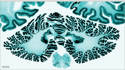


This summer, researchers from McGill University and the Montreal Neurological Institute (MNI) made headlines when they revealed the world’s highest resolution 3D model of the human brain in the June 20 issue of the journal Science. The model, which took nearly a decade to develop and is composed of one terabyte of data, has been dubbed the “BigBrain atlas.”
The atlas is a technical milestone in the field of human brain mapping because of its micron-scale resolution, which scientists achieved by using a microtome to meticulously slice the post-mortem brain of a 65-year-old woman into 7,404 slices—each 20 microns thin. This extensive process was led by Dr. Katrin Amunts, a researcher from Jülich, Germany. Her team stained and digitized each slice with a high-resolution scanner before sending the data to researchers in Montreal.
Dr. Alan Evans, co-developer of BigBrain and a McGill professor at the MNI, led the venture to statistically analyze each slice and reconstruct a comprehensive 3D model of the brain. He explained that this was the slowest part of the whole process.
“You have 7,404 slices of saran wrap all wrinkled, crinkled, ripped and torn, and somehow you have to take all of that data and stitch it back together again into a coherent three dimensional entity that is useful,” Evans said.
The reconstruction process was possible due to advanced statistical and computational tools. Evans’ team developed new software in order to clean, reconstruct, and register their data in 3D. One platform, called CBRAIN, allowed the group to manage their terabyte-sized data set and granted them access to the network of High-Performance Computing (HPC) facilities distributed across Canada. Without the use of these supercomputing facilities which allow large datasets to be processed on multiple computers in parallel, the project would have taken several more years to complete.
Researchers at the MNI can browse the BigBrain atlas on a large computer screen made up of four screens placed together. The database was also made publicly available online in order to promote research and the development of new neurological tools. Data is streamed on-demand to clients all over the world using a program called Atelier3D, which has previously been used to visualize works of art—including the Mona Lisa—in three dimensions.
“BigBrain […] can be used to redefine traditional brain maps that date back to the 20th century.”
The atlas will, in all likelihood, become known as the world’s most detailed reference brain. All brains are slightly different in shape and size, so neuroscientists use reference brains to compare data collected from different individuals on a standard template. Current templates, however, were acquired using Magnetic Resonance Imaging (MRI) and are limited in their resolution to the millimeter scale. This means that once zoomed in, each pixel will be equal to 1 x 1 x 1 mm of real life brain space. If the same is done with the BigBrain atlas, however, the pixel dimensions are 20 x 20 x 20 microns, which is smaller than the diameter of a strand of hair. This level of resolution allows neuroscientists to visualize brain structures at a near-cellular level.
 The advantage of having such a high-resolution mapping template is that data from a wide range of sources can be integrated and modeled at a a highly detailed level. For instance, molecular, genetic, and cytochemical data can be modeled in addition to low-resolution data acquired by modalities such as Positron Emission Tomography (PET) and functional MRI (fMRI), which acquire functional information about the brain as opposed to structural information.
Erin Mazerolle, a post-doctoral fellow in the McConnell Brain Imaging Centre at the MNI who uses MRI data in her research, explained the benefit of having a high-resolution brain template.
“It’s important for us to consider that the structures and functions we observe at the scale of millimeters with MRI actually result from much smaller structures,” she said. “BigBrain is an important step towards linking MRI findings to the microscopic scale, so that we can start to appreciate the underlying complexity that is otherwise not accessible with MRI.”
Another significant implication of the BigBrain atlas is that it can be used to redefine traditional brain maps that date back to the 20th century, such as the Brodmann atlas. This atlas, which was published in 1909 by German anatomist Korbinian Brodmann, is still the most widely used method of delineating the human cortex. However, it was limited by the technology of the era and was therefore based on properties of cells that could be seen through a microscope. The BigBrain atlas, in comparison, could be used to develop any number of brain maps based on structural or functional criterion extracted using computational methods.
Evans said he was pleasantly surprised by the feedback from the neurosurgical community. He described the reaction of Dr. William Feindel, a 95-year-old pioneer in the field of neurosurgery based in Montreal, when he first saw ‘BigBrain.’
“He was sitting in front of the screen exploring the amygdala and the hippocampus and saying he had never conceived of being able to do this because he was used to looking at a two-dimensional plate of the hippocampus,” Evans explained. “Now he’s moving backwards and forwards and up and down and it was delightful to see the reaction of someone of his stature.”
Feindel explained that the BigBrain could eventually aid neurosurgeons by allowing them to precisely implant depth electrodes, which are small electrodes used to record electrical activity from the brain to localize the focal point of a seizure or stimulate the brain to provide therapy for conditions such as Parkinson’s disease or clinical depression. Currently, neurosurgeons use a stereotaxic atlas—a mathematical map of the brain—as a guide when implanting electrodes. However, stereotaxic atlases are not three-dimensional.
“You have three dimensions available, but not simultaneously, so you look at a horizontal section of the brain or a sagittal section or a coronal section, but you can only do that separately. With […] BigBrain you can see them all together at the same time, so you have a confluence of information that you just can’t get on the other atlases. That’s why [this technology] represents a big advance.”
Since the BigBrain atlas was created using a single brain, as opposed to other reference brains that were created by averaging together hundreds of brain maps, a commonly asked question is whether it can be used to capture differences between brains. Evans responded that with the high-performance computational strategies possible today, the atlas can be warped into any statistical brain space.

“You can superimpose it on the average brain so that it is sitting in a statistical space that represents a population, but it retains the high-resolution detail that’s in the brain. You can get the best of both worlds at that point.”
Evans mentioned that there is still work to be done in the continuous refinement and spatial registration of the data.
“When you look at the BigBrain data set at the level of MRI, it looks fantastic. When you go in finer, you start to see the imperfections in the alignment of the individual slices.” These imperfections were caused by rips and tears that occurred during slicing as well as differences in the amount of staining across slices.
Evans’ group will continue to work on improving their current data set while they begin collecting data for future brain atlases. Potential projects would be to create 3D brain atlases for different types of brains such as a male brain, a young brain, and a diseased brain. There is also the potential to create a BigBrain atlas demonstrating the white matter tracts of the brain. Although the first data set took a decade to produce, future data sets will be finished much quicker because the technologies required to do so are now in place.
The BigBrain atlas is part of Europe’s Human Brain Project, a €7 billion venture to model the brain. Although the Human Brain Project predates Obama’s Brain Research through Advancing Innovative Neurotechnologies (BRAIN) initiative, both projects will take advantage of the latest computing technologies to collect and integrate data about the human brain, and are indicative of a growing trend in neuroscience in which large-scale supercomputing has become indispensible to research. As new technologies continue to be developed, BigBrain will only become more useful as a tool for neurosurgery, teaching and research. For now, it represents a major technical achievement in its own right, and a giant step forward in the quest to model and simulate the human brain.

Wow! I want to see more interesting stuff like this in the Tribune. Well written 🙂