
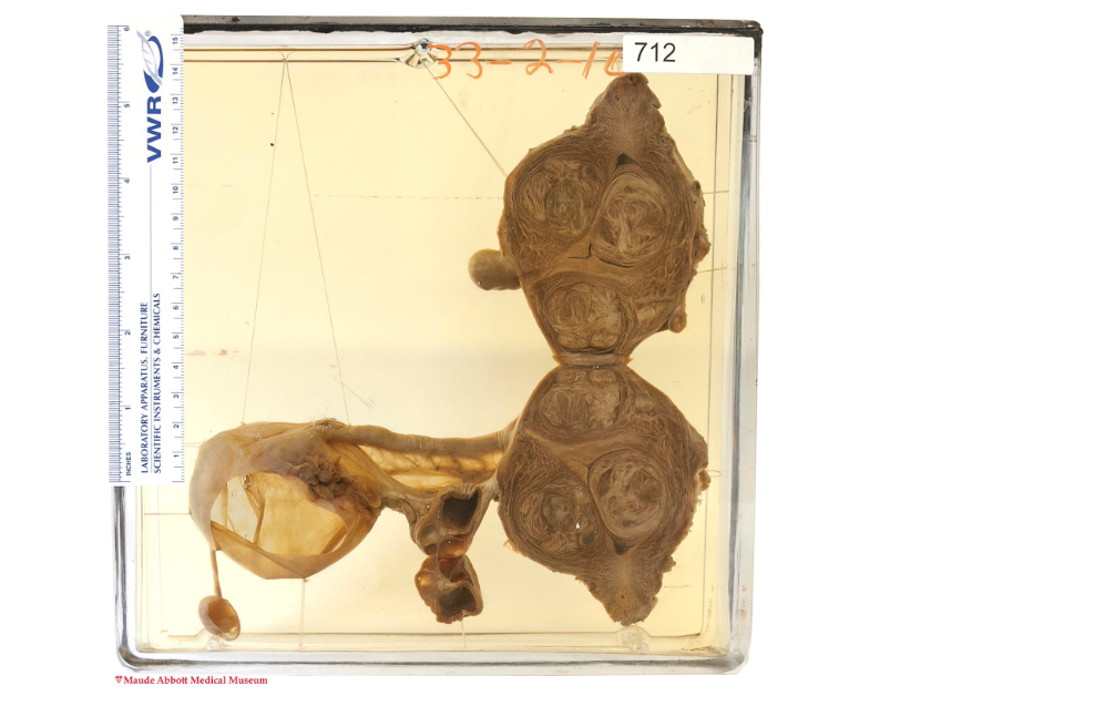

At its annual ‘Immortalizing the Mortal’ event in November 2019, the Maude Abbott Medical Museum revealed its chosen specimen: A uterus with large leiomyoma (or fibroids), benign tumours that occur when smooth tissue or connective tissue grows in the uterus. The event, which uses medical specimens to inspire artists and celebrate the artistic prowess of the participants, aims to explore the intersection of art and science to enable mortals to be immortalized through art focussing on their mystic medical histories. The specimen, which should be ‘artistically clothed with flesh,’ may be expressed through poetry, sketch, photography, or any other medium.
The Maude Abbott Medical Museum opened around the same time as the founding of the Montreal General Hospital in 1822 and the Montreal Medical Institute in 1823. The Holmes heart, acquired by McGill’s first Dean of Medicine Dr. Andrew Holmes in 1824, is the museum’s oldest and most recognizable specimen. In 1899, the museum was curated by Maude Abbott, the first woman to graduate from McGill with a Bachelor of Arts and the first woman to be admitted into McGill’s Faculty Club. After being denied entry into McGill’s medical school, Abbott went on to transform the medical museum into one of the best teaching museums in North America.
Richard Fraser, a pathologist at the McGill University Health Centre and curator of the museum, supervised the event. Fraser explained that the museum uses the specimens in the way that they were traditionally meant to be used, for analysis and medical diagnosis. However, hosting a creative event uncovers another side of the specimen. His vast knowledge on the topic allows event guests to learn about both pathology at McGill and the history of the field.
Pathology is a type of medical science in which scientists study surgically removed organs and tissues to diagnose disease. There are three different kinds of pathology: Cytopathology, molecular, and surgical pathology. Cytopathology is used to diagnose diseases such as cancer at the cellular level, while in molecular pathology, scientists study the molecules within the organs, tissues, and bodily fluids of the subject to diagnose and classify disease, such as cancer. For the 2019 specimen, participants applied surgical pathology, which involves examining tissues with the naked eye to provide a definitive diagnosis of the disease.
“The idea was to have an exercise whereby we get a specimen [that] clearly shows pathology but to treat it in a way that brings out the different aspects of the specimen,” Fraser said. “In other words, […] we take a structure […] and then find just the shapes or colours and the way it’s lined up […] or to try and look at the specimen from the point of view of the person that it came from.”
Fraser asked event participants to describe the specimen but not give it a diagnosis, before guessing what part of the body it was.
“I see circles and it’s symmetrical on both sides,” Henriette, a first-year Medicine student, said. “From what I can see it’s quite even, but there’s like a piece here that’s missing.”
Henriette guessed that the specimen was a brain, after which Fraser revealed the actual name.
“In fact, it’s a uterus, so you’re at the wrong end of the body,” Fraser said. “That’s an ovary, that sac, so this is an ovarian cyst, and the uterus has this whirl structure to it.”
The event staff hope that fellow participants will be similarly inspired by the shape or story of the specimen. All of the submissions of art will be presented at the wine and cheese on Jan. 17 from 5:00 to 7:00 p.m.