




For some individuals, recovering from COVID-19 is only the beginning of a longer battle with persistent symptoms. The World Health Organization estimates that 10 to 20 per cent of people infected with COVID-19 develop long COVID (LC), a condition marked by more than 200 symptoms that can emerge and persist months after initial infection. Yet despite the prevalence of LC, there is still no diagnostic test or validated biomarker that can identify the condition, forcing clinicians to rely largely on patients’ self-reported symptoms.
In a recently published paper in the Journal of Clinical Neuroscience, a team working with Monserrat Casado Sánchez—as she completed her PhD at McGill’s Brain Research and Imaging for Neurorehabilitation Lab (BRAIN) under the supervision of Marie-Hélène Boudrais—identified measurable differences between brain activity in people with LC versus those without LC. As people with LC report experiencing fatigue, brain fog, and post-exertional malaise (PEM), the researchers concluded that these patterns warrant further investigation as potential biomarkers for LC.
The BRAIN researchers were motivated to investigate LC because of their concerns about the limited scientific understanding of LC’s genesis and its effects on patients.
“There were two main things that were really worrying [about long COVID]. One, that symptoms were very debilitating, and two, that [its] origin was seriously misunderstood,” Casado Sánchez said in an interview with The Tribune.
Brain imaging studies have shown that LC leaves physical traces on the brain, including reduced grey matter volume and weakened thalamus-to-motor-region connections. Researchers have linked these changes with symptoms such as fatigue, impaired attention and memory, and slower processing speed. Less is known, however, about how these changes in brain activity are directly linked to long COVID symptoms.
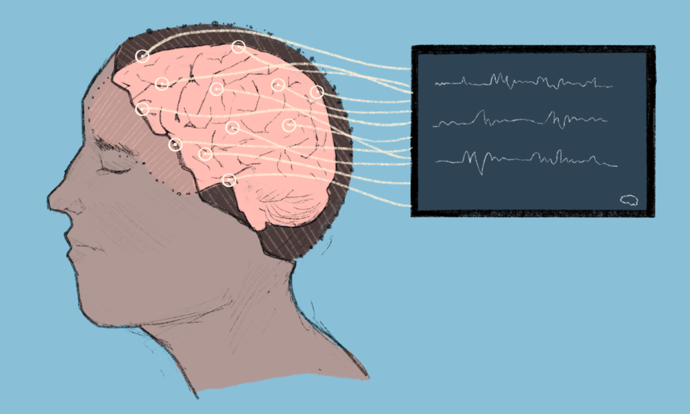
To investigate this connection, McGill researchers from BRAIN studied 20 right-handed adults with LC who reported fatigue, brain fog, and PEM, comparing them with 20 age- and sex-matched healthy control subjects. First, participants completed three validated symptom questionnaires designed to assess the presence and severity of their LC-related symptoms. Then, days or weeks later, the researchers recorded all subjects’ electroencephalogram (EEG) activity over the left motor and somatosensory regions of the brain during both rest and sustained handgrip contractions, measured using a handgrip dynamometer.
The BRAIN team focused on two measures: post-movement beta rebound (PMBR)—a burst of activity after movement—and alpha central frequency—the pace of brain waves at rest. A smaller beta rebound may reflect differences in how the brain regulates activity after movement, while a slower alpha rhythm may reflect differences in how it functions at rest.
Compared with healthy controls, participants with LC showed reduced PMBR following handgrip task and a slower alpha central frequency at rest. These changes correspond to specific LC symptoms: Participants with more severe brain fog tended to have smaller PMBRs, while those with greater fatigue tended to have slower alpha frequencies.
Since both measures correlated with participants’ reported symptoms, the findings suggest that, in people with LC, the brain regions involved in movement and sensation may have difficulty regulating their activity.
Casado Sánchez and her colleagues suggest that the findings could reflect that LC causes a disruption in the balance between excitatory and inhibitory brain signals, possibly involving neurotransmitters: though they remain uncertain of the underlying mechanism.
However, these associations alone do not definitively establish that altered brain activity causes fatigue or brain fog in patients with LC.
“It is super important to remind everyone and to remember that correlation does not equal causation [….] This was a small sample size and it was an exploratory study, so we cannot draw any hard conclusions to what these correlations mean,” Casado Sánchez cautioned.
Furthermore, similar alpha- and beta-band abnormalities have been reported across conditions including Parkinson’s disease, fibromyalgia, and myalgic encephalomyelitis/chronic fatigue syndrome, suggesting that these EEG features may reflect a broader response to chronic fatigue-related states rather than being unique to LC. The researchers suggest that these EEG features could eventually contribute to the development of objective biomarkers for LC, although further research is needed to determine whether they can reliably distinguish LC from other conditions.
“[We now] need validation in larger, more diverse groups, alongside longitudinal studies,” Casado Sánchez said.